Oral Leukoplakia. The importance of Early Diagnosis and periodic control with a Specialist
What is Oral Leukoplakia?
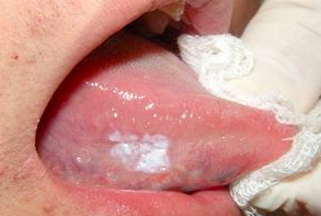
Leukoplakia is a clinical term that describes a predominantly white lesion of the oral mucosa, which cannot be scraped off and cannot be classified as any other entity.
The importance of oral leukoplakia is that it is the most frequent precancerous lesion of the oral mucosa, that is, leukoplakia has a proven risk of transforming into a cancer and the early diagnosis of this pathology is fundamental.
Tobacco use is the most important etiological factor known to date for the development of oral leukoplakia, although on some less frequent occasions leukoplasias may appear in non-smoking patients, and it is thought that some viruses might be involved.

The whitish coloration of leukoplasias is due to changes in thickness and keratinization of the epithelium.
Oral Leukoplasms, after microscopic study, are classified into 5 stages, according to the degree of epithelial alteration:
Simple Keratosis, Hyperplastic Leukoplakia, Leukoplakia with Dysplasia (with different grades), Carcinoma “in situ” and Carcinoma Invasor.
A different variant of Oral Leukoplakia is Hairy Leukoplakia, which appears on the lateral edges of the tongue, in a patient with AIDS, transplanted or with immunosuppression. These leukoplakias are caused by the Epstein Barr virus.
At Davalos and Balboa Dental Clinic, we are specialists in Oral Medicine. Come and see all your questions.
Dr. Balboa, our specialist in this area, recommends a general review of the mucous membranes annually, within the framework of the annual oral health review.


